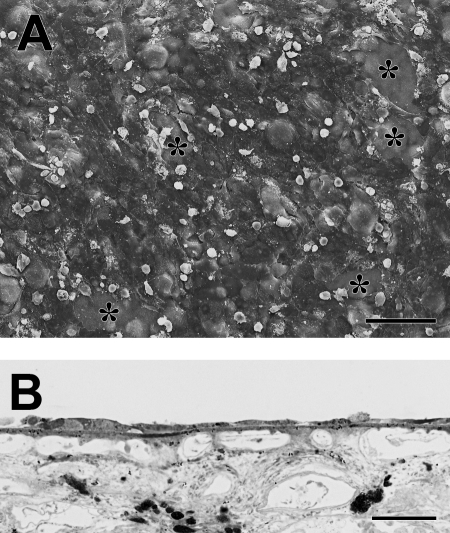
Figure 10.
Morphology of fRPE on submacular Bruch's membrane after 21 days in organ culture (donor age 69 years, same donor as Fig. 9A, 9B). (A) fRPE show more resurfacing of the explant than that observed by hES-RPE on the fellow explant. Cells on the incompletely resurfaced explant are very flat and highly variable in size. Large defects in cell coverage are indicated by asterisks (ND, 16.81 ± 0.39). (B) LM of the explant shows the variability in cellular morphology. Scale bar: (A) 100 μm; (B) 30 μm. Toluidine blue staining.

