Figure 6.
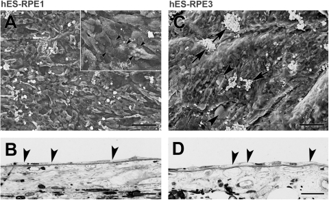
Morphology of hES-RPE on equatorial Bruch's membrane from a 59-year-old donor after 21 days in organ culture. (A) hES-RPE1 (ND, 6.10 ± 0.30). Cells incompletely resurface the explant with a mixture of large, elongate cells and enlarged, polymorphic cells. Inset: cells resurfacing the explant have smooth surfaces with no or few apical processes. At this magnification, small and large (arrow) vacuoles can be seen within many of the cells. The ICL is evident in regions with small defects in cell coverage (arrowheads point to some of the defects). (B) LM of the explant shows flattened, elongate cells on Bruch's membrane. Arrowheads point to part of flattened cells with vacuoles or loss of cytoplasm. (C) hES-RPE3 (ND, 15.44 ± 0.58). Cells almost completely resurfaced this explant with a few small defects in coverage (arrowheads). Cells are highly variable in size with varying degrees of vacuole formation. Rounded supernumerary cells are present on the surface of the monolayer (arrows). (D) LM shows hES-RPE3 resurfaced the explant with a monolayer of cells. Similar to the cells in B, vacuole formation and loss of cytoplasm are found in several of the cells (arrowheads). Scale bar: (A, C) 100 μm; (A, inset) 20 μm; (B, D) 30 μm. Toluidine blue staining.

