Figure 7.
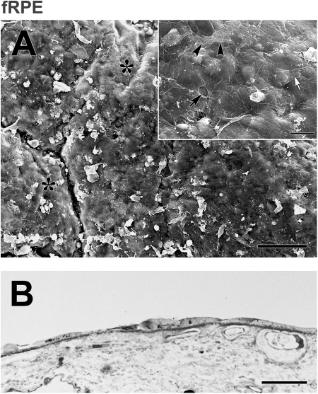
Morphology of fRPE on equatorial Bruch's membrane from a 59-year-old donor after 21 days in organ culture (same donor as Fig. 6). (A) fRPE incompletely resurfaced the explant with confluent patches of large, flat cells. Areas not fully resurfaced expose the inner collagenous layer (asterisks). Cell remnants are present (white debris; ND, 10.26 ± 0.41). Inset: The flattened appearance and smooth surfaces of the fRPE are more evident at this magnification. Unlike hES-RPE (Fig. 6), few vacuoles are present. A small intercellular gap is present (arrow). Cell extensions are not uncommon (white arrow points to lamellipodia extending over an adjacent cell). Arrowheads point to the border of a cell with a perforated cell membrane. (B) LM of the explant illustrates the variability in size and shape of fRPE. Scale bar: (A) 100 μm; (A, inset) 20 μm; (B) 30 μm. Toluidine blue staining.

