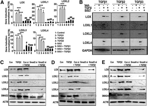
Figure 7.
Smad2, -3, and -4 inhibition blocked TGFβ-induction of the LOXs. Treatment of TM cells with the Smad3 inhibitor SIS3 blocked TGFβ1, -2, and -3 induction of LOX/LOXL mRNA (A) and protein (B) expression. (A) qRT-PCR analysis of the TGFβ1, -2, and -3 induction of the LOXs in the presence of a specific inhibitor of Smad3 (SIS3). qRT-PCR results represent the ratio of induction of LOX/LOXL genes normalized to ACTB, the housekeeping gene, in TGFβ-treated samples compared with the controls (triplicates of three TM cell strains). One-way ANOVA was used for statistical analyses. **,##0.0001 < P < 0.01; ***,###P < 0.0001. #Differences between TGFβ1, -2, and -3 samples versus TGFβ+inhibitor samples; *differences between TGFβ1, -2, and -3–treated versus untreated cells. (B) Western immunoblots of LOX/LOXL proteins after pretreatment with SIS3 followed by TGFβ treatment. Immunoblots are representative of three different TM cell strains treated with 5 ng/mL of TGFβ1, -2, and -3 for 48 hours along with 10 μM of SIS3. GAPDH was used as loading control. Untreated and DMSO-treated cells served as negative controls. (C, D, E) Western immunoblots of LOX/LOXL proteins in TM cells pretreated with Smad2 (C), -3 (D), or -4 (E) siRNAs followed by TGFβ2 treatment. Control cells were transfected with nontargeting siRNA. Immunoblots are representative of results from two TM cell lines. Each Smad siRNA not only knocked down its target protein, but also suppressed the TGFβ2 induction of LOX/LOX proteins.

