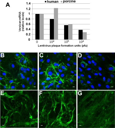
Figure 6.
Lentiviral shRNA silencing of versican in TM cells. (A) The indicated dilutions of versican shRNA lentivirus were added to porcine and human TM cells in culture, and versican mRNA was assessed by qRT-PCR 48 hours later. Values represent the ratio relative to mock-infected TM cells. (B–G) Immunofluorescence of porcine (B–D) and human (E–G) TM cells in culture using a versican monoclonal antibody for (B, E) mock-infected cells, (C, F) control lentivirus-infected cells, and (D, G) versican shRNA lentivirus-infected cells. Immunofluorescence was performed 72 hours after infection. DAPI was used to stain nuclei (blue). Scale bars, 10 μm.

