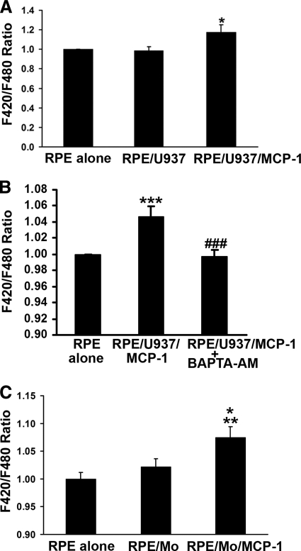
Figure 1.
Activated monocytes stimulate Ca2+ signals in RPE cells. (A) RPE cells were preloaded with 10 μM fluorescent Ca2+ indicator dye (Fura red-AM; Molecular Probes), and fluorescence intensities were measured from RPE cells exposed to control medium (RPE alone), U937 cells (RPE/U937), or MCP-1–activated U937 cells (RPE/U937/MCP-1). (B) Fluorescent Ca2+ indicator dye–preloaded RPE cells were preincubated with or without 10 μM BAPTA-AM for 30 minutes, and then exposed to control medium (RPE alone) or MCP-1–activated U937 cells in the presence (RPE/U937/MCP-1+BAPTA-AM) or absence (RPE/U937/MCP-1) of BAPTA-AM. Fluorescence intensities were measured. (C) Fluorescent Ca2+ indicator dye–preloaded RPE cells were exposed to control medium (RPE alone), monocytes freshly isolated from human peripheral blood (RPE/Mo), or MCP-1–activated monocytes (RPE/Mo/MCP-1). Data represent the mean ± SEM (n = 3–20). *P < 0.05, **P < 0.01, ***P < 0.001, compared to control or RPE/Mo; ###P < 0.001, compared to RPE/U937/MCP-1.

