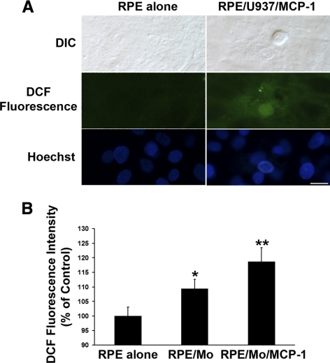
Figure 2.
Activated monocytes induce ROS production in RPE cells. (A) RPE cells were preloaded with 5 μM CM-H2DCFDA, and then exposed to control medium (RPE alone), or MCP-1–activated U937 cells (RPE/U937/MCP-1) for 1 hour. Differential interference contrast (DIC) and fluorescence images of intracellular ROS deposition were displayed. Scale bar, 20 μm. (B) RPE cells were preloaded with 5 μM CM-H2DCFDA, and then exposed to control medium (RPE alone), monocytes (RPE/Mo), or MCP-1–activated monocytes (RPE/Mo/MCP-1) for 1 hour. Intracellular ROS production was measured with a fluorometer (FlexStation Scanning Fluorometer; Molecular Devices). Data are presented as mean ± SEM (n = 3). *P < 0.05, **P < 0.01, compared with RPE alone (control).

