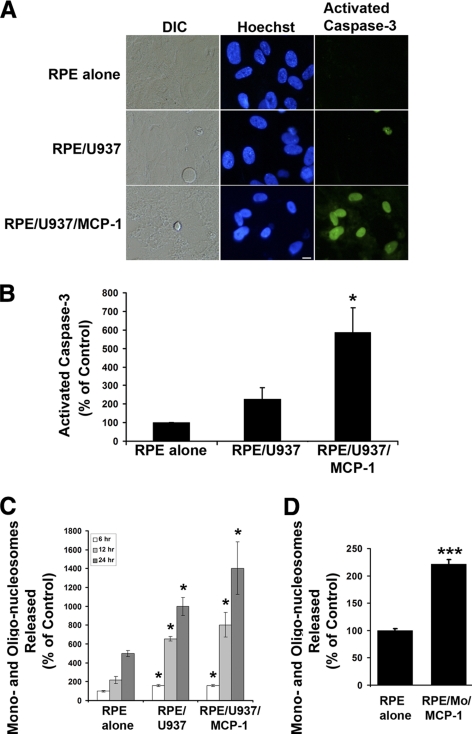
Figure 3.
Activated monocytes induce apoptosis in RPE cells. (A) Differential interference contrast (DIC) and fluorescence images of cells stained with Hoechst 33342 and caspase-3 substrate (NucView 488; Biotium, Inc.) in RPE cells exposed control medium (RPE alone), unstimulated U937 cells (RPE/U937), or MCP-1–activated U937 cells (RPE/U937/MCP-1). Scale bar, 10 μm. (B) A quantitative analysis of activated caspase-3–positive RPE cells. Activated caspase-3–positive RPE cells were determined after staining with caspase-3 substrate. (C) RPE cells were treated as described in (A) for 6, 12, and 24 hours. DNA fragmentation or released mono- and oligonucleosomes were measured by a cell death detection ELISA. (D) RPE cells were treated with MCP-1–activated freshly isolated human monocytes (RPE/Mo/MCP-1) for 24 hours. DNA fragmentation or released mono- and oligonucleosomes were measured by a cell death detection ELISA. Data are presented as mean ± SEM (n = 3–6). *P < 0.05, ***P < 0.001, compared with RPE alone (control).

