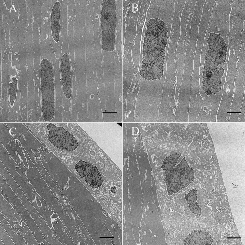
Figure 3.
Electron microscopy photo of lenses from WT (FVB/N strain) and CRYGC5bpd mice of 5 weeks. Lens fiber cells from CRYGC5bpd mice (B) appear edematous with prominent nucleoli compared with WT control mice (A). Moreover, lens epithelial cells in CRYGC5bpd are swollen compared with WT (D and C, respectively). Scale bar, 2 μm.

