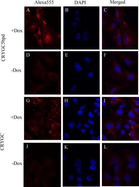
Figure 5.
Fluorescence confocal microscopic pictures. The red fluorescence in the first column is Alexa 555 Fluor from the secondary antibody, whereas the nuclei of cells were counterstained with DAPI and are seen blue in second column. The third column shows merged images of both stains. The top two rows show the Tet-on advanced HeLa cells transfected with CRYGC5bpd. γC-crystallins were expressed by adding Dox in (A–C). (D–F) show the transfected cells growing without Dox. The bottom two rows show the WT CRYGC transfectants, Dox-induced expression (G–I) and without Dox γC-crystallins were not expressed (J–L). There is no significant difference between WT and mutant crystallins, although the levels of WT CRYGC appear to be somewhat higher than those of CRYGC5bpd in these experiments.

