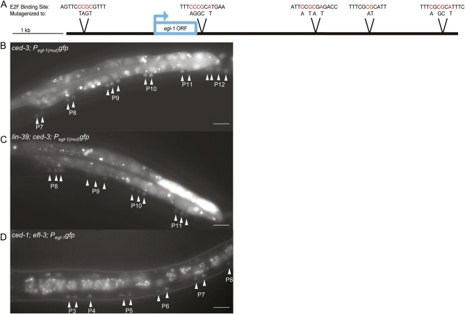
Figure 5 .
A repressive E2F binds Pegl-1. (A) Five E2F consensus sites were identified in Pegl-1. At each site, the nucleotides shown in red were mutated to abolish E2F binding. All five mutated sites were combined into one construct to give Pegl-1(mut). (B) Loss of E2F-binding sites in Pegl-1 results in ectopic egl-1 expression in midbody P3–P8 and posterior P9–P12 lineages. Mutations in Pegl-1gfp induce a doublet pattern of neurons in the midbody and an increase of neurons in the posterior expressing egl-1. (C) In lin-39; Pegl-1(mut)gfp animals, GFP is expressed in a triplet pattern in the midbody and most posterior lineages. (D) efl-3(null); Pegl-1gfp animals exhibit ectopic egl-1 expression. Scale bars, 10 μm. Animals in B and C are homozygous for ced-3, and the animal in D is homozygous for ced-1.

