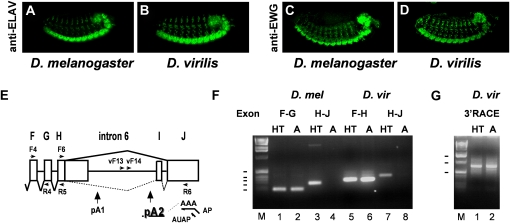
Figure 1 .
ELAV and EWG expression is conserved in D. virilis. (A–D) Expression of ELAV (A and B) and EWG (C and D) in D. melanogaster embryos (A and C) and D. virilis embryos (B and D). (E) Schematic of ewg intron 6 from D. melanogaster. ELAV-regulated splicing of intron 6 is shown as a solid line, and non-neuronal splicing of introns 6a and 6b is shown by a dashed line. The ELAV-regulated poly(A) site is underlined. Schematic position of primers used in both species for PCR or for 3′ RACE in D. virilis (vF13 and vF14) are indicated by arrows. (F and G) RT-PCR of ewg in D. melanogaster and D. virilis. Splicing of intron 6 is confined to the neuron-rich head/thorax (HT) and reduced in the neuron-poor abdomens (“A” in F and G) in both species as shown in F. ewg cDNAs from exons F and G in D. melanogaster, exons F and H in D. virilis, and exons H–J in both species were amplified with primers F4 and R4 or R5, and F6 and R6, respectively. 3′ RACE confirms a single intronic poly(A) site (pA) in intron 6 in D. virilis as shown in G. 3′ RACE of cDNAs in D. virilis was done with nested primers vF13 and vF14, and the return primer AUAP in two reactions on AP reverse-transcribed RNA. Molecular weight markers (M) are indicated on the left in F (100–500 bp) and G (0.5, 1, and 1.5 kb).

