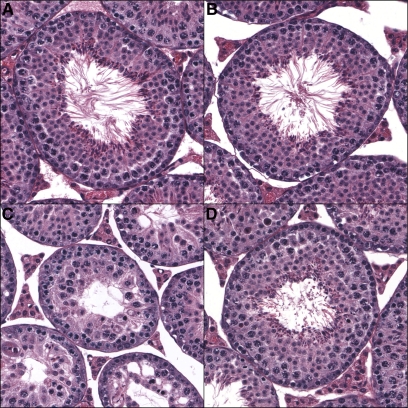
Figure 3 .
Stage VII seminiferous tubules. Cross sections from (A) M. m. domesticusWSB, (B) M. m. musculusPWD, (C) (M. m. musculusPWD × M. m. domesticusWSB)F1, and (D) (M. m. domesticusWSB × M. m. musculusPWD)F1 males. In the sterile F1 direction (M. m. musculusPWD × M. m. domesticusWSB)F1, there is a large reduction in post-meiotic cells, reducing the overall area of the tubule. All images are at ×200 magnification.

