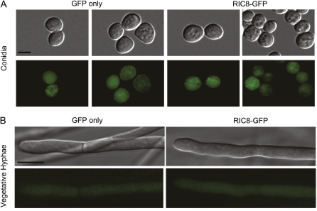
Figure 4 .
Localization of RIC8-GFP in Neurospora. Cultures were grown as indicated in Materials and Methods. Images were obtained using an Olympus IX71 microscope with a QIClickTM digital CCD camera and analyzed using Metamorph software. (A) DIC and GFP fluorescence micrographs showing localization of RIC8-GFP in conidia. Bar, 5 μm. (B) DIC and GFP fluorescence micrographs showing localization of RIC8-GFP in vegetative hyphae. For both A and B, GFP fluorescence images for a wild-type strain without a GFP construct were completely black. Bar, 10 μm.

