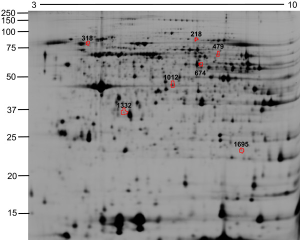
Figure 1.
2D-DIGE of peripheral neutrophils from COPD patients and healthy controls. Neutrophils were isolated from the peripheral blood and protein lysates for 2D-DIGE were prepared. Proteins were focused on 24 cm pH 3-10 NL IEF strips and separated by 12% SDS-PAGE. Representative image of the biological variation analysis (BVA) software is shown. The differentially regulated proteins are indicated by the red spot boundaries.

