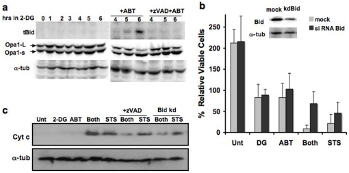
Figure 4. The Bid-based feedback loop is required for efficient 2DG-ABT induced apoptosis.
a, Bid is activated during 2DG-ABT induced apoptosis. Western blots of PPC-1 cell lysates treated with 10 mM 2DG for 0–6 hours (left panel), with either 1 µM ABT-263 alone or ABT-263+10 µM z-VAD for the last 3 hours (right panel), probed with the anti-tBid antibody (upper panels), anti-Opa1 antibody (middle panels), and anti-alpha-tubulins (lower panels). Note: the loss of Opa-1-L was used as a marker for remodeled mitochondrial cristae during cytochrome c release from the mitochondria [34]. b, The effects of Bid depletion on 2DG-AB-induced apoptosis. Insert: western blots of PPC-1 cell lysates, mock transfected (left lane) or transfected with Bid siRNA (right lane). Bid (+) and Bid (−) PPC-1 cells were treated with either 2DG, ABT-263 (1 µM), both, 10 µM ABT-263, or left untreated for 24 hours. The viable cells were counted and the increase/decrease over the input numbers were graphed. c, In PPC-1 cells either pre-treated with 2DG for 3 hours or without pre-treatment, then 1 µM ABT-263 or 1 µM STS was added for three hours, and cytosolic fractions were analyzed by western blots. In parallel experiments, Bid-depleted PPC-1 cells were treated with the combination of 2DG and ABT-263 or with STS and analyzed (designated as Bid kd, last 2 lanes). Cells were harvested 3 hour after ABT/STS treatment, and cytosolic fractions were analyzed with western blots.

