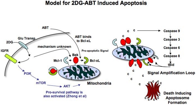
Figure 6. Model of 2DG-ABT Induced Apoptosis.
a, 2DG is specifically taken up by cancer cells through a glucose transporter. Once inside, it is phosphorylated and becomes a hexokinase inhibitor. At the same time, it activates several signal transduction cascades. One of them is IGFR-PI3K-mTOR-AKT pro-survival pathway. Through another cascade activation, Bak-Mcl-1 association is lost. When ABT is added 3 hours later, it can bind to Bcl-xL and disrupts its association with Bak. b, Freed Bak oligomerizes, releasing a small amount of cytochrome c into the cytosol. It activates cascades of caspases 9-3-6-8 and caspase 8 cleaves Bid, generating BH3 only tBid protein. Since tBid can dissociate Bak-Mcl-1 and Bak-Bcl-xL association, it induces mitochondria outermembrane pore formation through which more cytochrome can be released. Released cytochrome c recruits Apaf1, caspase 9 and dATP, forming a “death executioner” apotosome complex.

