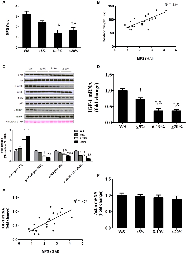
Figure 1. Muscle protein synthesis and IGF-1/mTOR signaling are reduced during the progression of cachexia in ApcMin/+ mice.
Protein synthesis and IGF-1 expression were measured in ApcMin/+ mice during the progression of cachexia. A) Myofibrillar protein synthesis. B) Correlation between muscle weights and protein synthesis. C) Upper: representative western blot of phosphorylated and total forms of Akt (Ser 473), mTOR (Ser2448), p70S6k (Thr389) and 4EBP-1 (Thr37/46). Lower: The ratio of phosphorylated and total Akt, mTOR, p70 and 4EBP1 in the gastrocnemius muscle normalized to the WS group. D) IGF-1 expression and E) correlation between IGF-1 gene expression and protein synthesis. F) Skeletal alpha actin mRNA expression. Values are means ± SE. Significance was set at p<0.05. † Signifies different from WS mice. & Signifies difference from mice with ≤5% body weight loss. $ Signifies difference from mice with 6–19% body weight loss. WS, weight stable.

