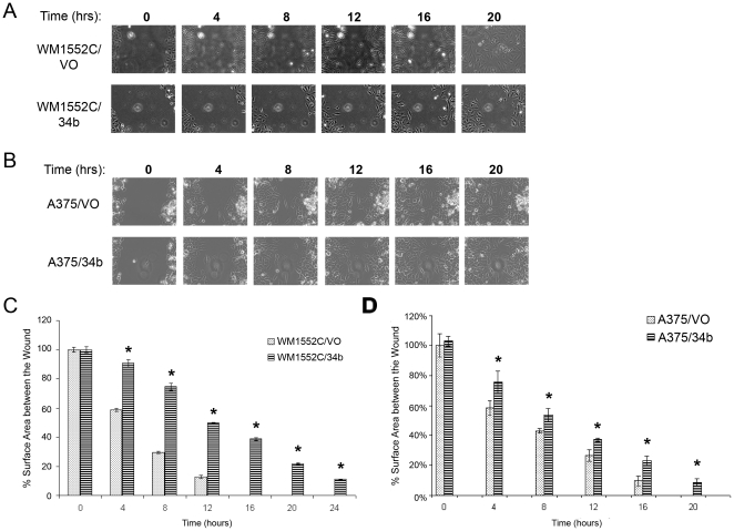
Figure 6. Effect of miR-34b on cell migration.
Wound healing assay for melanoma cells. A) and B) Representative images of WM1552C/VO and WM1552C/34b cells or A375/VO and A375/34b cells. Images were taken between 0 and 20 hrs after scratch formation. C) and D) Quantitation of experiments shown in (A) and (B), performed in triplicate. The percent surface area between the wounds was calculated using NIS elements software. Each time point was compared with the 0 hr time point of the respective cell line. Asterisks indicate statistical significance by Kruskal Wallis nonparametric test, P<0.005.

