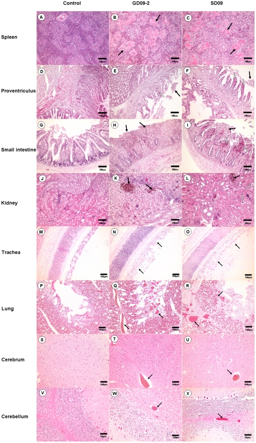
Figure 3. Histopathology on tissues from 1-week-old chickens infected with NDV GD09-2 or SD09 (H&E).
B and C: amalgamation of collapsed cell and inflammatory exudates created the homogeneous and pink-staining appearance in the white pulps of spleens (black arrow); E and F: dropout and necrosis of the mucosal epithelia in the proventriculus (black arrow); H and I: dropout of epithelium and numerous inflammatory cell infiltration in the small intestine (black arrow); K and L: congestion (black arrow) or glomerulus atrophy (white arrow) in the kidneys; N and O: dropout and necrosis of mucous epithelial cells in the trachea (black arrow); Q and R: congestion and hemorrhage in the lung (black arrow); T and U: venous congestion in the cerebrum (black arrow); W and X: venous congestion in the cerebellum (black arrow); A, D, G, J, M, P, S and V: Corresponding control tissues. Scale bar = 50 µm in cerebellum or 100 µm in other tissues.

