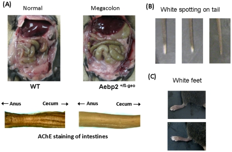
Figure 3. Phenotypes of the Aebp2 +/β-Geo mice.
(A) Comparison of internal organs between the wild-type (WT) and Aebp2+/β-Geo mice (upper panel). Some of the Aebp2+/β-Geo mice display an enlarged green-colored colon (Megacolon), which is easily detectible as compared to the normal-size colon from the wild-type mice. Acetylcholinesterase staining further indicates that the Aebp2+/β-Geo mice have much less ganglion cells in the intestinal section between the anus and cecum than the wild-type mice. The ganglion cells are shown as brown thin fibers on the surface of the intestines (lower panel). Some of the Aebp2+/β-Geo mice also display white spotting at the tail tip (B) as well as the toes (C).

