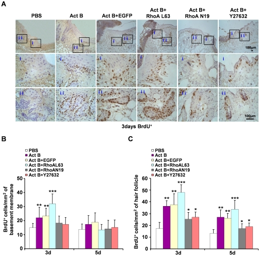
Figure 5. Activation of RhoA contributes to the proliferation of epithelium keratinocytes and hair follicle cells during wound healing.
Twenty four mice were randomly assigned to four groups: Act B+EGFP (n = 6), Act B+RhoAL63 (n = 6), Act B+RhoAN19 (n = 6) and Act B+Y27632 (n = 6). (A) Mice were treated with Act B+EGFP, Act B+RhoAL63, Act B+RhoAN19 or Act B+Y27632 after wound creation. Immunohistochemistry was performed on the indicated day after treatment. BrdU-positive cells on epithelium (i) and hair follicle (ii), pictures were taken at 100× magnification, and corresponding magnification pictures i and ii were taken at 400× magnification, Scale bars: 100 µm. Data were quantified in three independent experiments and the number of BrdU-positive cells per mm2 in epithelium (B) and hair follicle (C) is presented. *P<0.05 compared with PBS control. **P<0.05 compared with PBS, Act B+RhoAN19 and Act B+Y27632 groups. ***P<0.05 compared with PBS, Act B, Act B+EGFP, Act B+RhoAN19 and Act B+Y27632 groups.

