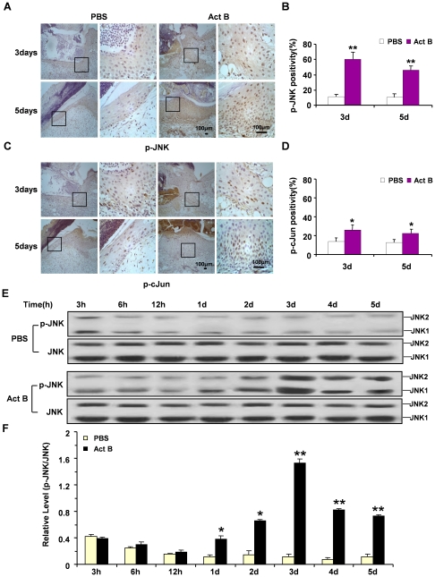
Figure 6. Activin B induced activation of JNK and cJun in wounded skin.
Twelve mice were randomly assigned to PBS group and Act B group (n = 6). Immunohistochemistry indicated increased numbers of p-JNK-positive (A) and p-cJun-positive cells (C) at the wounded skin on day 3 and day 5 after activin B treatment, as compared with PBS control. Enlarged images of the boxed area were presented on the right side. Scale bars: 100 µm. Quantified percentages of p-JNK and p-cJun immunopositive cells are presented in (B) and (D) respectively. (E, F) Western blotting showed the level of JNK phosphorylation reached the highest value on day 3 and higher than PBS group on day 1 to day 5. 3 h, 6 h, 12 h: 3, 6 and 12 hours after wound. *P<0.05, **P<0.01, compared with PBS control.

