
Fig. 2.
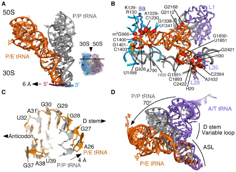
Conformation of tRNA in the P/E hybrid state. (A) Movement of P/E tRNA and mRNA towards the E site when compared to P/P tRNA and mRNA. The direction of view is shown to the right. (B) View of mRNA and P/E tRNA interactions with the 30S subunit P site and 50S subunit E site. Residues that contact mRNA (gold) and P/E tRNA (red) are shown. Colors for the ribosome, mRNA and tRNA as in Fig. 1. (C) View of the P/E tRNA ASL/D stem junction (orange). P/P tRNA (grey) is shown for comparison, with an arrow indicating the widening of the helix major groove. (D) Comparison of ASL/D stem junctions between P/E tRNA (orange), P/P tRNA (grey), and A/T tRNA (purple). A/T tRNA structure is a homology model adapted from (12, 21). The bending angle for the A/T to P/E conformational change (70°) is shown.