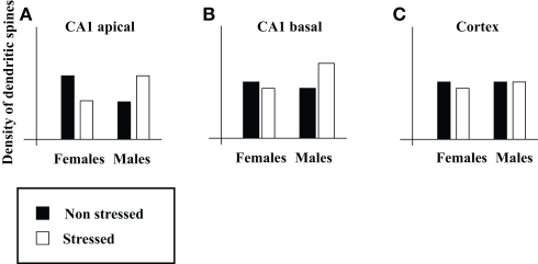
Figure 1.
Illustration of the effects of acute stress on sex differences in density of dendritic spines. Illustrations of (A,B) the density of the apical (A) and basal (B) dendritic spines on pyramidal neurons in CA1, and (C) the density of dendritic spines in somatosensory cortex (on the basis of Figure 4 in Shors et al., 2001).

