
Figure 1c:
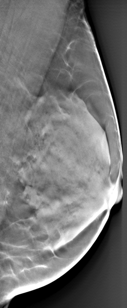
Differences in parenchymal tissue texture visualization in a 56-year-old woman on (a) a digital mammogram, where parenchymal tissue layers are superimposed, and (b, c) DBT images, where the superficial skin layer, in which the skin pore texture is seen in b, is separated from the deeper fibroglandular tissue layers, as seen in c.