
Figure 1:
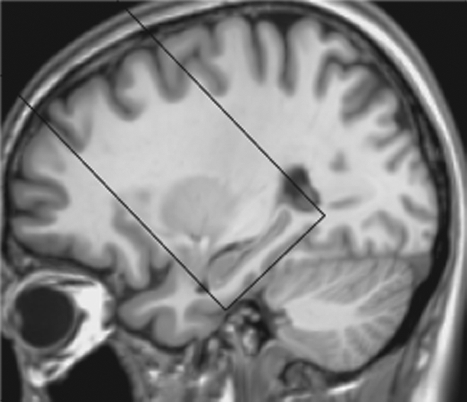
Image shows ASL perfusion imaging slab position. The oblique coronal ASL imaging sections were positioned to cover the head of the hippocampus, with the inferior edge of the imaging slab parallel to the longitudinal anteroposterior axis of the hippocampus and tangent to the gyrus of the temporal lobe, by using T1-weighted high-spatial-resolution anatomic images for accurate reference.