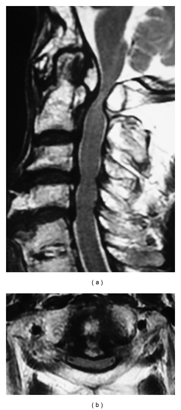
Figure 3.

Preoperative MRI on T2-weighted image. (a) Sagittal plane. The spinal cord is compressed at the level of C1, C3-4, and C4-5, with high intensities within the spinal cord at C1 level. (b) Axial plane. The spinal cord is compressed by the mass behind dens and posterior arch of the atlas.
