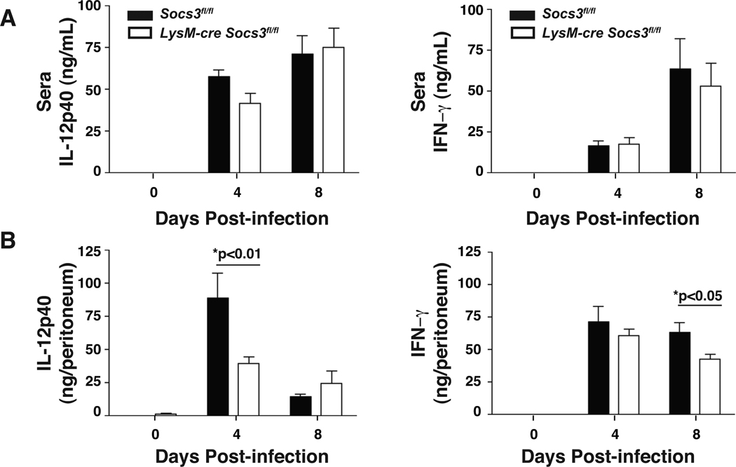
Figure 5. Infected LysM-cre Socs3fl/fl mice show impaired IL-12 production in the peritoneal cavity at day 4.
Peritoneal lavage fluid from infected LysM-cre Socs3fl/fl mice contains a lower concentration of IL-12p40 at day 4 that corresponds to a similar trend in sera obtained from blood. Although IFN-γ production appears normal at day 4, a diminished IFN-γ response was observed at day 8 in the peritoneal lavage fluid. (A,B) Fluid from intraperitoneal lavage of PECs and serum of infected mice at days 4 and 8 were collected and analyzed by ELISA for IL-12p40 and IFN-γ. PEC fluid results are presented as ng/peritoneum which is calculated by multiplying the ELISA concentration result (ng/mL) by the mL quantity of peritoneal lavage fluid (PBS) recovered following centrifugation. One-Way ANOVA followed by student’s t test was used to determine statistical significance. Error bars +/− SD. Results are representative of two independent experiments.

