Figure 1.
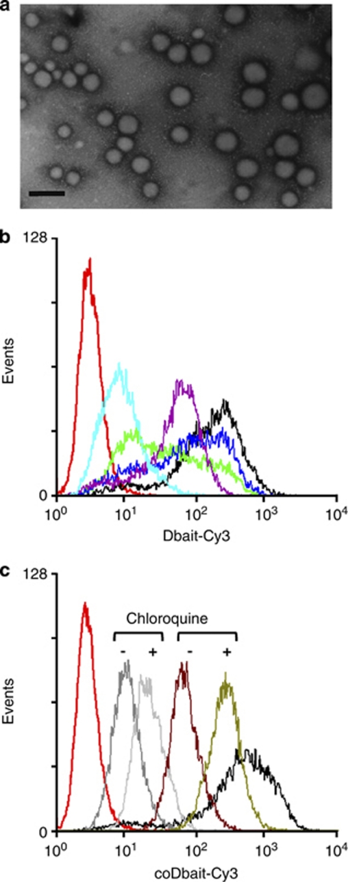
Cellular uptake of formulated Dbait. (a) Microscopic analyses of Dbait complexes with polyethylenimine (PEI)11k. (b and c) Flow cytometric analyses of cellular uptake were performed 5 h after beginning treatment for various transfection conditions. (b) 1.6 μg ml−1 Dbait-cy3 (red), Dbait-cy3 with Superfect (black), bPEI25K (purple), PEI11K (green), PEI22K (blue), electroporation (light blue); (c) Dbait-cy3 with Superfect (black), 1.6 μg ml−1 coDbait-cy3 (grey), with and without chloroquine (CQ) treatment (light grey) before transfection, 21 μg ml−1 coDbait-cy3 without CQ (brown) and with CQ (green).

