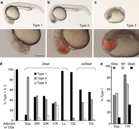
Figure 4.
Phenotypes 24 h after Dbait injection into the extracellular space of cell stage 1 K zebrafish embryos. (a–c) Lateral views anterior to the left of zebrafish embryos, 24 h after Dbait-cy3+polyethylenimine (PEI) injection (2–5 nl) at the animal pole of cell stage 1 K zebrafish embryos: top panel, bright field view; bottom panel, 2 × magnification of the head region with epifluorescence overlay showing in red Dbait-cy3, scale bar 250 μm. (a) Type 1 phenotype indistinguishable from non-injected (not shown). (b) Type 2 mild phenotype with extensive cell death in the head region. (c) Type 3 strong teratogenesis and widespread cell death. (d) Histogram showing the percentage of the three phenotypic classes depending on the adjuvant. More than 100 embryos were analyzed for each condition. Sup: Superfect; 25, 22, 11 K PEI of the corresponding size; Lu, Lutrol; CQ, chloroquine; ‘−', without adjuvant. (e) Histogram showing the percentage of the animals with type 1 phenotype after injections of nanoparticules formed with 8H, 64ss or Dbait32H and Superfect or PEI11K. The Dbait32H, and the 8H and 64ss inactive nanoparticules, were used at equivalent adjuvant concentrations.

