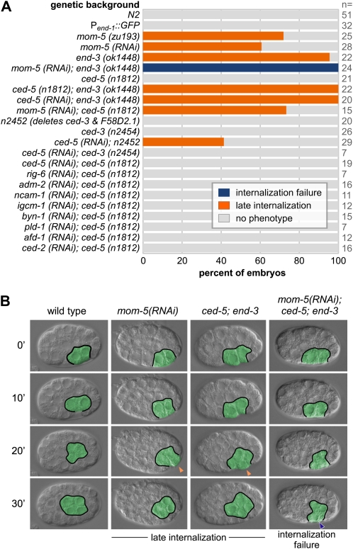
Figure 1 .
Enhancement of subtle gastrulation defects. (A) Gastrulation defects in various mutants and/or from injected dsRNAs. (B) Four-dimensional (4D) DIC microscopy of four backgrounds with time on the left from MSa/p cell division. E lineage cells are outlined and pseudocolored in green. Late internalization at the 4E stage (orange arrowheads) and internalization failure (blue arrowhead) are indicated. “No phenotype” indicates that endodermal precursors became internalized at the 2E stage, as in wild-type embryos. Scale: C. elegans embryos are ∼50 µm long.

