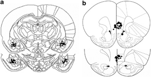
Figure 1.
Schematic drawing indicating electrode tip positions within the medial prefrontal cortex (mPFC) and the basolateral amygdala (BLA). Shown is a coronal view of (a) the mPFC and (b) the BLA at position 3.20 mm and 2.70 mm anterior to the bregma, and at −3.14 mm and −3.30 mm posterior to the bregma, respectively. Adapted with permission from Elsevier 1986, Paxinos and Watson, 1986.

