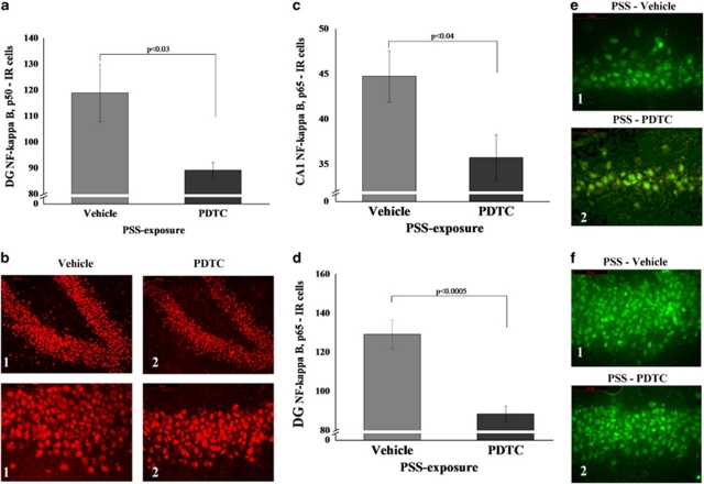
Figure 12.
Effect of early post-stressor intervention with PDTC on NF-κB p50 and p65 immunoreactivity in the hippocampus. (a) The quantitative analysis of p50 subunit immunostaining in the DG area of exposed rats treated with PDTC or vehicle. (b) Representative photographs of NF-κB p50 immunoreactivity in the DG of exposed animals treated with PDTC (2) or vehicle (1). Photographs were acquired at × 20 (Scale bar, 100 μm) and × 40 magnification (Scale bar, 50 μm). The cells in red were p50 positive. (c) The quantitative analysis of p65 subunit immunostaining in the CA1 (c) and DG (d) areas of exposed rats treated with PDTC or vehicle. (e) Representative photographs of NF-κB p65 immunoreactivity in the CA1 (e) and DG (f) of exposed animals treated with PDTC (2) or vehicle (1). Photographs were acquired at × 40 magnification. Scale bar, 50 μm. The cells in green were p65 positive. Administration of high-dose corticosterone immediately post exposure significantly decreased expression of NF-κB p50 and p65 subunits in the DG when compared with exposed animals treated with vehicle. All data represent group mean±SEM. DG, dentate gyrus; CA1, cornu ammonis 1; EBR, extreme behavioral response; MBR, minimal behavioral response; PBR, partial behavioral response; PSS, predator scent stress. The color reproduction of this figure is available at the Neuropsychopharmacology journal online.

