
Figure 3.
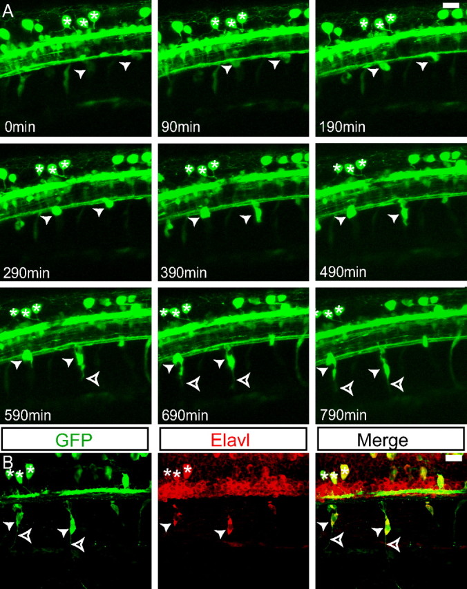
GFP-positive cells become DRG neurons. A, Individual frames of z-projections of a confocal time-lapse movie of a Tg(neurog1:EGFP) from 26 to 39 hpf. GFP-positive DRG precursor cells become visible at the ventral edge of the spinal cord (arrowheads). The same embryo is shown in supplemental Movie 1 (available at www.jneurosci.org as supplemental material). As the cells mature, they send out projections (open arrowheads). A group of RB neurons is maintained throughout the duration of the imaging (asterisks) and serves as a landmark for registration. B, Immunolabeling of the embryo at 40 hpf. The DRG cells from A are labeled with both anti-GFP and anti-Elavl antibodies, indicating that they have differentiated as neurons (arrowheads). Axons extend ventrally from the DRG neurons (open arrowheads). The group of RB neurons remains identifiable (asterisks). Scale bars, 20 μm.