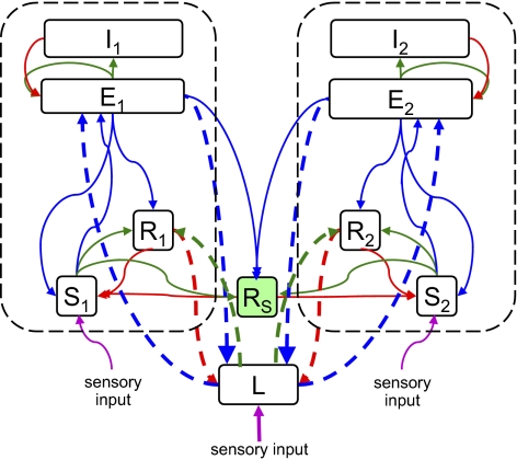
Fig. 3.
Addition of shared thalamic excitation to the model of Fig. 1. The heavy dashed arrows are connections involving L, which represents a thalamic nucleus that projects to two cortical regions. Thin arrows represent connections already present in the model of Fig. 1; see Fig. 1 caption for color conventions.

