
Fig. 6.

Identification of fluctuations of coherence modes in EEG data. (A) Spectrograms and coherograms from model simulations (Left) and surrogate data (Right). Below the coherogram is the time course of the first principal component and the histogram of these values. (B) A parallel analysis of patient EEG recordings at F7 and F8 (Laplacian derivation) under baseline conditions (Left) and following zolpidem administration (Right). The vertical bars superimposed on the spectrograms and coherograms indicate the subdivision of the data into epochs. Calibration bars apply to all horizontal axes in A and B and to the vertical axis for the first principal component; full time axes are 300 s (A), 93 s (B, Left), and 240 s (B, Right). C summarizes the evidence for multimodality in the coherences measured between multiple electrode pairs (significance level indicated by color: red,  ; orange,
  ; yellow,
 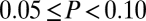 ; L, R, A, and P indicate left, right, anterior, and posterior, respectively; see key for 10–20 system electrode designations) and 18FDG-PET images of metabolic activity [standardized uptake values (ref. 30) indicated by color]. Note that following zolpidem, multimodality appears in the frontal channels, along with an increase in frontal metabolic activity.