Fig. 4.
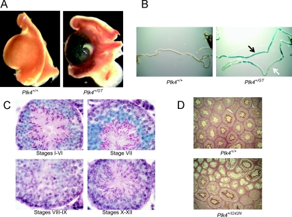
Plk4 expression in the testis. A, Whole mount testis LacZ staining from E18.5 Plk4+/GT mice. B, Whole mount LacZ staining of seminiferous tubules dissected from testes of 6-wk-old Plk4+/GT mice. Black arrow indicates areas of high expression. White arrow indicates areas without expression. C, Sections from 6-wk-old Plk4+/GT mice stained with LacZ and PAS-H. D, Immunohistochemical staining of testes sections from 6-wk-old Plk4+/+ and Plk4+/I242N mice with an anti-PLK4 antibody.

