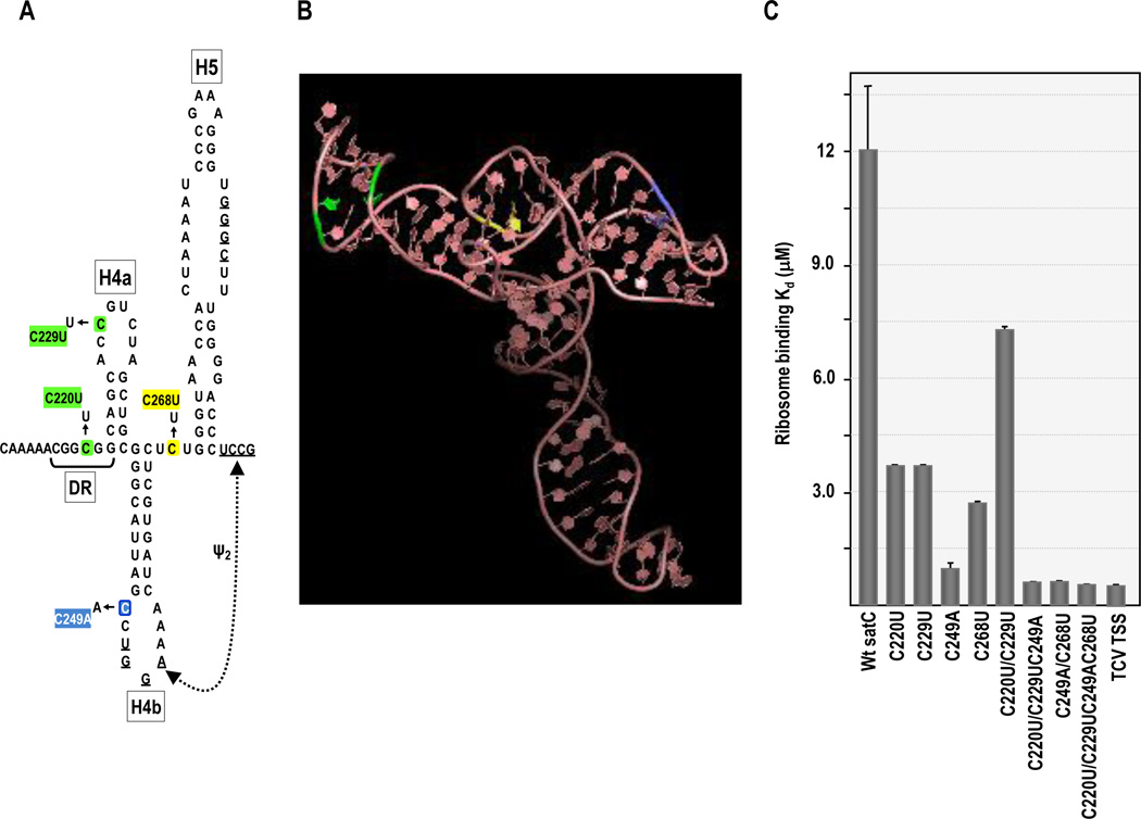
Fig. 2.
Ribosome binding of satC mutants containing TCV TSS sequence. (A) SatC TSS analogous region. Location of nucleotide differences is indicated. Colors of particular alterations correspond with their location in the TSS structure. (B) 3D structure of the TCV TSS determined by NMR and SAXS [small angle x-ray scattering; (Zuo et al., 2010)]. Locations of nucleotides differences with satC are color coded as in (A). (C) 80S ribosome binding of satC mutants. Filter binding assays were performed in triplicate and standard error is given.

