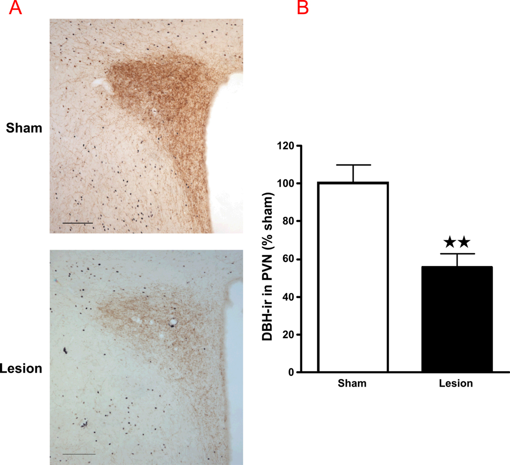
Figure 6.
(A) Anti-DBH-saporin lesions significantly decrease the number of DBH-positive fibers in the PVN, compared to sham-operated rats. Bright-field photographs through the PVN from sham controls (top) and lesion rats (bottom) challenged with alcohol. Images show a representation of the immunohistochemical procedures that stained DBH fibers in brown (scale bar = 200 µm). (B) The density of DBH-ir fibers in the PVN. Mean ± SEM density of DBH-ir fibers in the PVN of sham and lesion rats injected with alcohol. ***, P<0.001.

