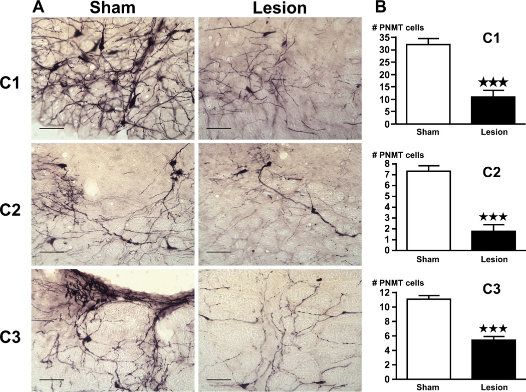
Figure 7.
(A) Lesions caused by the anti-DBH-saporin toxin significantly decreases the number of PNMT-positive cells in the C1–C3 cell groups, compared to rats pretreated with the vehicle. Bright-field photographs through the C1–C3 area of brain stem from sham controls (left) and lesion (right) rats challenged with alcohol. Images show a representation of the immunohistochemical techniques that stained PNMT-ir in purple (scale bar = 200 µm). (B) Cell counts were obtained for PNMT-ir positive cells in the C1, C2 and C3 area of the brain stem. Mean ± SEM levels of PNMT-ir cells in the C1–C3 area of sham and lesion rats injected with alcohol. ***, P<0.001.

