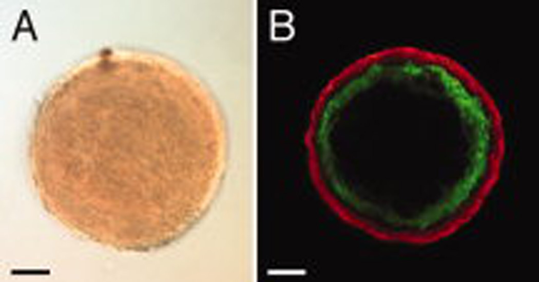
Figure 1. Uniluminal vascular spheroids have an outer SMC layer expressing SM22α and an inner cavity lined by ECs expressing CD34.
A, Light microscopic image of a uniluminal vascular spheroid generated in hanging drop culture. B, A single optical section from a laser scanning confocal microscopy (LSCM) z-series projection through the center of a uniluminal vascular spheroid that was immunolabeled with antibodies to CD34 (green) and SM22α (red). Bars equal 100µm.

