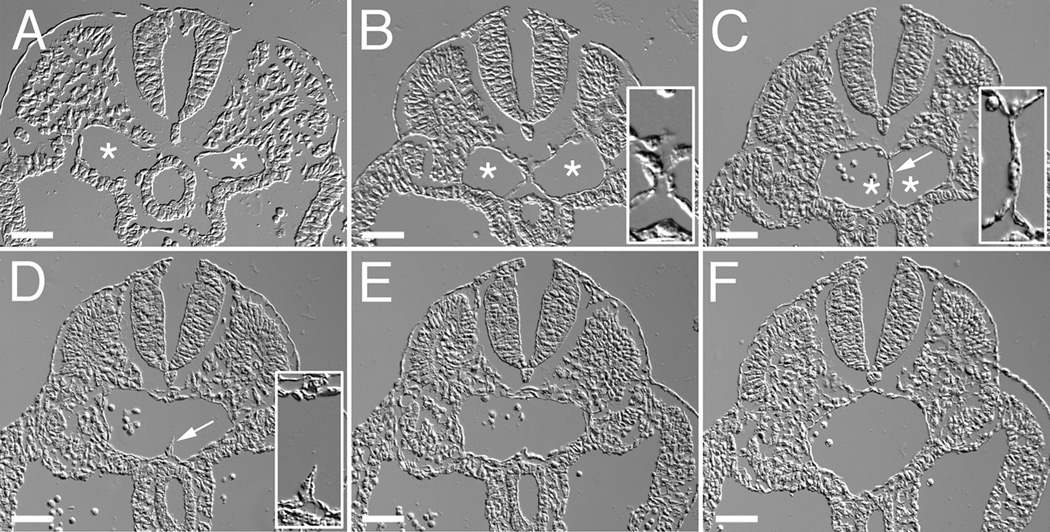
Figure 8. The aorta forms through the fusion of paired blood vessels that extend along the long axis of the embryo.
Shown are DIC images of a series cross-sections from positions along the posterior-anterior axis of an E9.5 mouse embryo. Sections are arranged in a posterior (A) to anterior (F) direction. Asterisks indicate the lumens of the paired aortae (A–C). Arrow in C shows the cellular septum separating the two aortae. Arrow in D shows a remnant of the cellular septum. Insets in panels B and C show high magnification views of the interface region between fusing dorsal aortae. Inset in panel D shows a high magnification view of the remnant of the cellular septum in the dorsal aorta. Bars equal 50µm.

