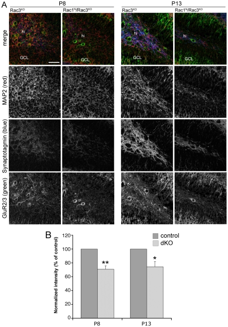
Figure 2. Synaptogenesis is affected in the hilus of double knockout mice.
P8 and P13 sagittal sections of dorsal hippocampi were immunostained for the indicated antigens. (A) Confocal images of MAP2 (red), GluR2/3 (green), and synaptotagmin 1 (blue) immunoreactivity within the hilus of control Rac3KO and Rac1N/Rac3KO double knockout littermates. The three markers are expressed in the hilus of P8 and P13 control animals, and they are strongly reduced in the hilus of P8 and P13 double KO mice, respectively. GCL, granule cell layer of the dentate gyrus; hi, hilus. Bar: 50 µm. (B) Quantification of synaptotagmin 1 fluorescence intensity in the hilar neuropil of Rac3KO (control) and Rac1N/Rac3KO (dKO) littermates at P8 and P13, respectively. The signal for synaptotagmin 1 was reduced in the double knockout mice, both at P8 and P13. Bars are average values from 13 (P8) or 10 (P13) sections from 3 mice per genotype. *P<0.05; **P<0.001.

