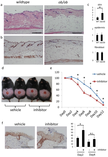
Figure 7. Selective 11β-HSD1 inhibitor enhance wound healing in ob/ob mice.
(a,b) Representative H&E staining (a) and 11β-HSD1 staining (b) of 6-week-old male wildtype and ob/ob mice. Bar = 50 µm. (c) The relative expressions of 11β-HSD1 in epidermis, fibroblasts, and whole skin extract of wildtype and ob/ob mice assessed by rtPCR. GAPDH was used as an internal control (P<0.05, Student's t-test). (d) Macroscopic view of wound healing on day 8. A 15-mm wound was created on the back of 6-week-old male ob/ob mice and wound closure was monitored with application of vehicle or 11β-HSD1 inhibitor every other day. (e) Reduction of wound area on days 2, 4, 6, 8, 10, and 12. The histograms indicate means and standard deviations for four mice in each group. An asterisk indicates a statistically significant difference (*P<0.05, Student's t-test). (f) Representative Ki-67 staining in day4 wound edge skin and the percentage of Ki-67 positive cells in day2 and day4 wound edge epidermis. Analyses were performed by counting the total number of basal cells and cells expressing nuclear Ki-67 stain. Bars indicate mean ± SD of vehicle-treated mice (n = 3) and inhibitor-treated mice (n = 3; *P<0.05, Student's t-test). Bar = 100 µm.

