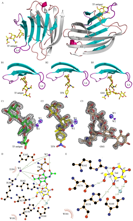
Figure 3. Overall structures of Gal-3 CRD complexed with TF antigen and its derivatives.
(A) Two Gal-3 CRD-TF complex molecules in an asymmetry unit. TF antigen bound to the CRD concave is shown in a stick model. (B) The ligands binding site in complexes Gal-3-TF antigen (B1), Gal-3-TFN (B2) and Gal-3-GM1 (B3), which show a conservative location at the concave formed by β-strands S4, S5, S6 and a loop L4 connecting S4, S5. (C) Fo-Fc electron density maps around TF (C1), TFN (C2) and GM1 (C3) with two conservative water molecules (contoured at 3.0 σ). (D, E) Interactions between Gal-3 CRD and TF antigen or TF moiety in TFN and GM1, in which the GalNAc and Gal moieties are shown in green and yellow, respectively.

