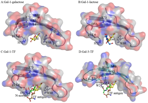
Figure 7. Structural comparison of the carbohydrate recognition pockets in Gal-1 and Gal-3 binding with different ligands.
(A, B) The concave carbohydrate binding pocket in Gal-1 shows Gal-1 recognizes and binds with galactose (Gal) or alternative lactose suitably. (C) The modeled TF antigen shows a serious spatial obstruction between its N-acetyl group and His52 on g1-L4. (D) The TF-binding pocket and the unique binding mode conserved in Gal-3 CRD. All ligands are shown in a ball-and-stick model and carbohydrate binding pockets are illustrated as surface drawing.

