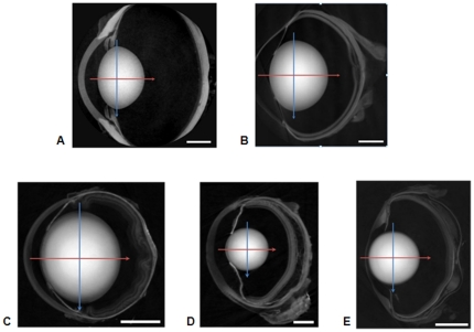
Figure 1. Images of a) porcine; b) ranine; c) murine; d) newt; e) piscine eyes in the sagittal plane.
The position of the equatorial plane is marked with the blue arrow and the optic axis, along which the sagittal refractive index profiles were measured, is marked with a red arrow. The scale bars in the right hand lower corner are equal to a) 4 mm (porcine); b) 2 mm (ranine); c) 1 mm (murine); d) 0.5 mm (newt); e) 1 mm (piscine).

