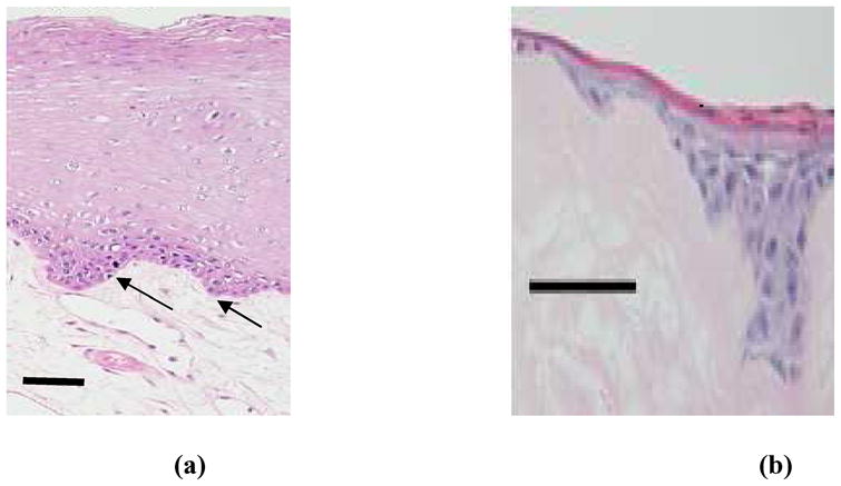
Figure 2.

Histology of natural oral mucosa tissue (a) and EVPOME device (b). In both cases, there is a basal layer of actively dividing and differentiating cells (arrows), followed by a uniformly keratinized surface (asterisk). Scale bars represent 100μm.
