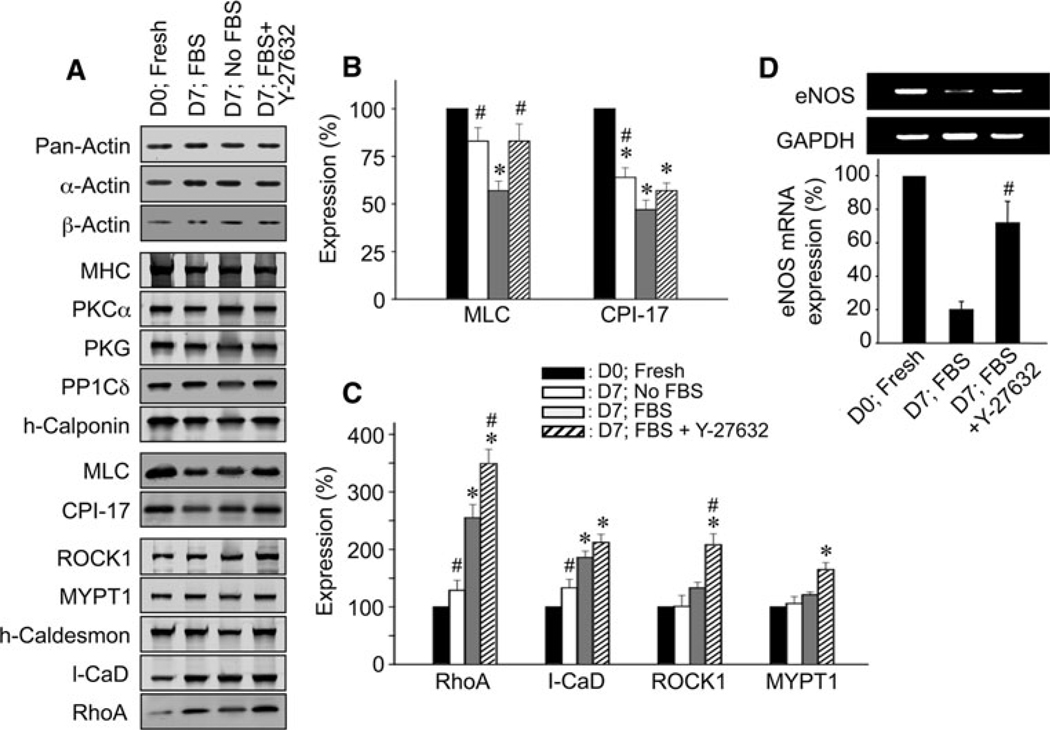
Fig. 2.
Changes in protein expression after 7 days of organ-culture and the effect of 10 µM Y-27632 in rabbit mesenteric artery. A Representative immunoblot images of various proteins from freshly isolated (D0; Fresh) and 7 days cultured arteries in FBS-free medium (D7; No FBS), and FBS-supplemented medium in the absence (D7; FBS) and presence of Y-27632 (D7; FBS + Y-27632). Total actin (pan-actin) contents under different conditions were matched. For actins, protein extracts diluted ten-fold were loaded. B and C Average expression levels of contractile/regulatory proteins (n = 4–6). D Representative RT–PCR images (upper) and a quantitative summary of eNOS mRNA expression (lower, n = 4). The symbols for statistical significance are defined as described in the legend of Fig. 1

